This leg carries a large proportion of a horse's weight but is not attached to the rest of the body by bone! This means that the leg (through the muscles) is able to absorb much of the concussive forces which would otherwise go straight to the spine. This lack of bony attachment is because the horse does not have a collar bone! There are a number of different bones which make up the foreleg. The bones are shaped to provide points for the attachment of muscles.
Is flat and shaped almost like a triangle as it is narrower at one end than the other. The outer surface of the scapula has a ridge in the centre (or slightly off centre) called the scapula spine, either side are muscles. The spine provides an attachment for the Trapezius muscle. The inner surface of the scapula is hollow to allow space for another muscle - the subscapularis. The Scapula also provides attachment for several other muscles including the Rhomboideous and the Triceps .
At the narrow, lower end the Scapula has a cavity (the glenoid cavity) which is where the head of the humerus sits. The upper end is attached to the thoracic vertebrae.
The shoulder joint is formed between the glenoid cavity and the head of the humerus. It is contained within a joint capsule as explained in my blog about joints. The humerus itself is one of the strongest bones in the body. The upper end is large with a convex head which fits into the cavity already mentioned. This bone again provides attachment for many muscles. The lower end has a groove which allows for the articulation (joining ) with the radius and ulna bones.
The Radius and Ulna
These bones are joined together with fibres when the horse is young but as the horse ages this turns to bone. This is unlike these bones in a human where the bones are able to move separately thus allowing the palm to be turned upwards. In most animals, including humans, the ulna is larger than the radius - in the horse the radius is the larger bone. The ulna does though have a large projection - the olecranon process. This is the bone that 'sticks' out on the horse's elbow!
The Elbow Joint is the hinge joint created between the Radius and Humerus.
The Carpus
This is the knee and there are usually 7 carpal bones (sometimes 8) which make up the carpus. These are mostly small squarish shaped bones with the exception of the accessory carpal bone which sticks out at the back providing the 'pulley block' for the leverage of the muscles.
Side view of the knee.
The Metacarpals
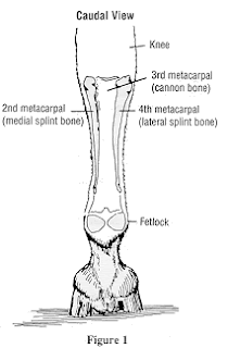
There are actually 3 of these; the main metacarpal which is capable of carrying a lot of weight and the 2 smaller splint bones. This large metacarpal (cannon bone) is the equivalent of a human middle finger, the -splint bones the index and ring fingers!
View from the back.
The Phalanges
The First Phalanx: (Long Pastern) articulates with the large metacarpal and the Second Phalanx. The top is grooved, this is where the cannon bone sits. The common digital extensor tendon attaches to a prominence at the front of the upper end of the long pastern and the superficial flexor tendon attaches at either side at the back of the lower end!
The Second Phalanx: (Short Pastern) articulates with the First Phalanx and the Third. The deep flexor tendon supports this bone at the back of the leg. As with the long pastern, the common digital extensor tendon also attaches to the front of the short pastern and the superficial flexor tendon to the back. The angle of this bone in the foot and the support of the deep flexor tendon work to lessen the concussive force which the bone is subjected to.
The Third Phalanx: (Pedal or Coffin Bone) is a similar shape to the hoof but only takes up a small area of the hoof itself. This bone articulates with the Second Phalanx at the top but also with the Navicular bone (distal sesamoid bone) at the back. The extensor process on the front of the upper end provides an attachment for the common digital extensor tendon. The deep flexor tendon attaches to the underneath edges of the bone by tendon in the shape of a fan. The digital or plantar cushion (see pictures below) provides a 'buffer' to the pedal bone and the deep flexor tendon against concussion. Underneath the cushion is the frog! Laminae help support this bone within the hoof.
The Proximal Sesamoid Bones: the 2 bones lie either side of the fetlock. They articulate with the cannon bone at its lower end. These bones act together to form a 'pulley block' for the deep digital flexor tendon. The bones are surrounded by ligaments to reduce the concussive forces which they have to endure.
The Distal Sesamoid Bone: is also known as the Navicular bone which lies behind the point where the Second and Third Phalanges meet. The deep digital flexor tendon runs over here (protected by a layer of fibrocartilage) as it goes to join the Pedal bone.
Next week: the hindleg.
Have you seen this week's video 'Christmas Treats' on my You Tube channel.
Horse Life and Love. Please check it out and SUBSCRIBE.
You can also follow me on Facebook and Instagram for updates on Chesney, Basil, Tommy and Daisy.
Until next time!
Jo


No comments:
Post a Comment